


Ultrasound
-
in Diagnostics
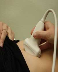
Ultrasound (ultrasound examination) is a procedure for examining the organs of the human body using ultrasound, which can penetrate living tissues. Ultrasound helps to observe and study the shape, position, structure, size and movement of various organs, as well as diagnose many diseases and monitor their development.
The "Alergologi" Medical Center offers to undergo an ultrasound examination on a modern "color apparatus" with the function of energy doppler.
Check the price
What is Doppler and why is it needed?
Doppler measures how sound waves bounce off moving objects. The computer processes the information and creates a two-dimensional color image that shows whether there are difficulties in the blood flow, for example, due to the deposition of cholesterol - atherosclerotic plaques.
Modern devices with continuous-wave, pulse-wave and color doppler combine the information obtained during all these types of research. In B-mode, you can see the structure of the vascular wall. Doppler shows how blood flows through the vessels and measures the speed of blood flow. Ultrasound can also be useful for determining the diameter of the vessel, as well as the degree of stenosis (blockage) of the blood vessel.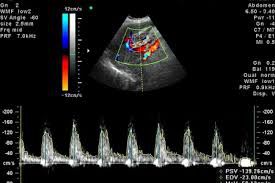
Traditional ultrasound uses painless sound waves, inaudible to the human ear, that bounce off blood vessels. Duplex ultrasound can use images that are color-coded to show the doctor where blood flow is severely blocked, as well as the speed and direction of blood flow. Your doctor may recommend a Doppler to help diagnose or investigate conditions that affect the blood vessels. These conditions include:
- atherosclerosis of carotid arteries;
- thrombosis of the veins of the lower and upper extremities;
- diseases of the arteries of the legs;
- diseases of the arteries of the hands;
- aorto-iliac occlusive diseases;
- varicose disease;
- aneurysms of large vessels of the abdominal cavity or extremities.